PCR vs qPCR Difference: Endpoint Versus Real-time Assays
The difference between PCR and qPCR is an assay-detection distinction that separates conventional endpoint product readout after cycling from real-time fluorescence measurement used to report Cq or Ct values. Endpoint PCR typically visualizes amplicons on a gel after the run. qPCR records signal every cycle so labs can quantify target when standards and efficiency checks are valid.
Molecular biologists use this comparison when deciding whether a cloning check, size-based genotype, or presence-absence test is enough, or whether relative or absolute quantification is required. Primer design, controls, and assay records change with that choice.
Core difference between PCR and qPCR
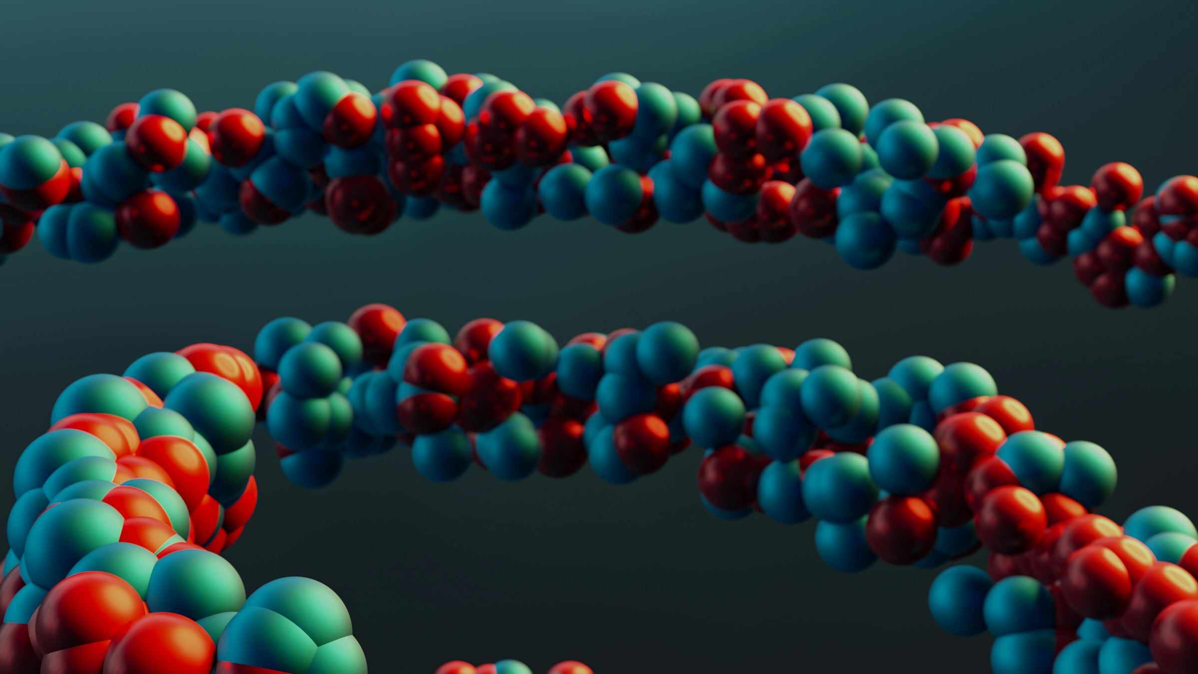
Both conventional PCR and qPCR amplify a DNA target with oligonucleotide primers, a thermostable polymerase, dNTPs, and a programmed thermal cycler. The shared chemistry is why the two methods are often discussed as if they were interchangeable. They are not interchangeable at the decision layer. Endpoint PCR asks whether a product of an expected size appeared after a fixed cycling program. qPCR asks how the fluorescence trace rose during those cycles, and whether that rise can be turned into a quantity.
In endpoint PCR, the reaction is typically opened after cycling and loaded on an agarose gel beside a DNA ladder. A band near the predicted length supports the claim that amplification occurred and that the product is consistent with the intended amplicon. Band presence does not prove sequence identity, frame, or the absence of a closely sized contaminant. Intensity on a stained gel is also a weak quantity. Saturation, loading differences, and stain binding prevent a gel from substituting for a calibrated standard curve.
In qPCR, also called real-time PCR, a fluorescence channel is read at a defined point in each cycle. An intercalating dye increases signal when double-stranded DNA accumulates. A sequence-specific hydrolysis or hybridization probe increases signal only when that third oligonucleotide engages the intended amplicon. Software then reports a quantification cycle, Cq, often still called Ct. Cq reports the cycle where the trace crosses a documented threshold. When the assay is behaving exponentially, a lower Cq indicates more starting target than a higher Cq in the same run.
Quantification is not granted by owning a real-time instrument. Relative quantification needs reference targets, matched sample quality, and a stated analysis model. Absolute quantification needs a standard of known copies and a curve that covers the working range of the samples. In both cases, amplification efficiency must be inspected rather than assumed. A well-behaved serial dilution supports efficiency review. A single Cq without context does not.
Comparison table: detection, output, and use cases
The table below compresses the PCR versus qPCR difference into the dimensions that change lab work: when product is observed, what number or image is stored, and which experimental question each format can answer.
| Decision dimension | Endpoint PCR | qPCR (real-time PCR) |
|---|---|---|
| Detection timing | After cycling ends | Fluorescence recorded each cycle |
| Typical output | Gel band size versus a ladder | Amplification trace plus Cq or Ct |
| Quantification | Not a reliable quantity from band brightness | Relative or absolute only when standards and efficiency are valid |
| Detection chemistry | No real-time reporter required | Intercalating dye or sequence-specific probe |
| Fits when | Cloning amplicon checks, size genotyping, simple presence or absence | Expression ratios, residual target, copy estimates from a standard curve |
| Core limitation | Size match is not sequence proof | Invalid Cq values without controls, a defined threshold, and efficiency review |
Read the table as a fit map, not a ranking. A cloning bench that only needs to confirm insert size should not be forced onto a quantification platform. A knockdown study that needs fold change should not treat a late gel band as a quantity.
When endpoint PCR is the right assay
Endpoint PCR remains the right assay when the scientific decision is categorical or size-based. Cloning workflows use it to confirm that a PCR fragment of the expected length was produced before ligation, Gibson assembly, or Golden Gate. A clean band near the map prediction is a useful gate. It is not a substitute for Sanger or NGS verification of junctions, tags, and reading frame after the clone exists.
Colony PCR is another endpoint job. Researchers screen colonies for insert presence, orientation, or cassette size, then advance only the colonies that match the expected pattern. The value is throughput and cheap exclusion of empty or wrong-size clones. The risk is treating a correctly sized colony PCR band as proof that the plasmid is the intended sequence. Point mutations, mixed colonies, and similar-length off-target inserts still pass a gel gate.
Genotyping by amplicon length also fits endpoint PCR. An insertion, deletion, or lox-flanked cassette that changes product size can be scored from a gel without converting the question into copies per microliter. Simple presence-or-absence tests follow the same logic: a pathogen-research screen, a mycoplasma check, or a barcode PCR may only need to know whether a product appeared with the expected controls. If the later decision is binary, quantification adds cost without changing the call.
Endpoint PCR also fits labs that lack an optical cycler, or that already have a standard thermocycler and gel documentation workflow. That is an operations reason, not a scientific ranking. The same lab should still move the question to qPCR when the notebook entry must report a ratio, a residual copy estimate, or a time-course of target abundance. Planning the reaction itself, including template input and cycling, is covered in Zettalab's guide on how to plan a PCR experiment.
When qPCR is required for quantification
qPCR is required when the experimental claim is quantitative. Relative quantification compares a gene of interest to one or more reference targets in the same cDNA or DNA preparation. The analysis is only as sound as the reference choice, the RNA integrity assumptions, and the demonstration that both assays amplify with comparable efficiency in the relevant range. If those conditions are not shown, a Cq difference is an observation, not a fold-change result.
Absolute quantification converts Cq into copies by reference to a standard curve. The standard must have a known identity and a stated copy or mass assignment. Samples should fall inside the curve, not below the last reliable point or above a saturated range. Extrapolation outside the standards turns Cq into a guess. Report the standard source, dilution series, and whether inhibition was checked, for example by dilution agreement or a spike-in, rather than a single undiluted well.
Chemistry choice changes what a Cq can mean. Dye-based assays report any double-stranded product, so primer-dimers and off-target amplicons can shift Cq and must be reviewed with melt profiles or gel spot checks during validation. Probe-based assays add sequence selectivity, but they also add oligo design burden and do not remove the need for no-template controls. Multiplex probe panels can quantify more than one target in one well only when competition, fluorescence crosstalk, and efficiency have been shown for that panel.
qPCR is the wrong default when the team only needs a size check or a yes-or-no cloning gate. It is also the wrong default when the sample set has no valid standard, no reference strategy, and no time to validate efficiency. In those conditions, a precise-looking Cq table can overstate certainty. Use qPCR when the question is abundance and the lab is prepared to document the calibration that makes abundance defensible.
Primer, control, and documentation implications
Primer design changes with the assay job. Cloning primers are often written to carry restriction sites, homology arms, or a distinctive amplicon length that is easy to score on a gel. qPCR primers are usually written for a shorter amplicon, limited secondary structure, and a single specific product. Reusing a long cloning primer pair in a real-time assay can lower efficiency and hide off-target products behind a late Cq. Design the oligo against the decision you will actually make.
When primers sit on a shared construct map, a molecular biology workspace such as ZettaGene can keep primer-binding coordinates attached to the same plasmid record used for later amplicon-size checks. That attachment matters because a renamed FASTA paste easily loses the intended start and end. ZettaGene is relevant here only as a place to review those primer-to-map attachments before oligos are ordered, not as a substitute for wet-lab validation.
Controls are not optional decoration. Endpoint PCR needs a no-template control to detect contamination or primer-dimer, a positive control of known size when a negative gel would otherwise be ambiguous, and a ladder that makes the size claim inspectable. qPCR needs those ideas in optical form: no-template wells, reference or standard samples, and, for RNA workflows, a no-reverse-transcriptase control so residual genomic DNA is not counted as transcript. Extraction or inhibition checks belong in the record when sample matrices vary.
Documentation should capture the assay type, primer sequences or IDs, master-mix lot, cycling program, detection chemistry, and the decision the run was meant to support. For qPCR, also store threshold-setting method, Cq values, melt or probe notes, standard identity, and the efficiency review that justified quantification. For endpoint PCR, store the expected size, gel image, and whether the next step is cloning, sequencing, or a simple pass-fail. Zettalab lab guides are useful when those fields need to sit beside the construct rather than in a disconnected spreadsheet.
FAQ
What is the difference between PCR and qPCR assays?
The difference is detection timing and the kind of claim the run can support. Endpoint PCR inspects product after cycling, typically as a gel band of an expected size. qPCR measures fluorescence during each cycle and reports a Cq or Ct value. Both assays still amplify DNA with primers and polymerase. Endpoint PCR fits size checks, colony screens, and presence-or-absence calls. qPCR fits abundance questions, but only after controls, a documented threshold, and an efficiency or standard-curve review. A bright gel band is not a copy number. A Cq without those checks is not a validated quantity either. Choose the format that matches the decision, then record the evidence that format can actually produce.
When should a lab use endpoint PCR instead of real-time qPCR?
Use endpoint PCR when the decision is whether a product of the expected size is present, not how many starting copies were in the tube. Cloning amplicon checks, colony PCR, and length-based genotyping are the common cases. Endpoint PCR also fits when the lab needs a fast exclusion gate before sequencing, or when no optical cycler is available. Do not use a gel as a substitute for qPCR just to save time if the paper or notebook will later state fold change or residual copies. In that case the missing calibration will have to be repeated. Keep endpoint PCR for categorical or size decisions, and move to qPCR when others will reuse an abundance result.
How do Ct values change qPCR data interpretation?
Ct, now often reported as Cq, is the cycle where the fluorescence trace crosses a stated threshold. In a valid exponential assay, a lower Cq means more starting target than a higher Cq in the same run and chemistry. Interpretation changes as soon as threshold setting, baseline, inhibition, or efficiency is poor. Then a Cq shift can reflect technical artifact rather than biology. Compare Cq values only inside a documented analysis method. For relative work, interpret Cq differences against reference targets and shown efficiencies. For absolute work, interpret Cq against a standard curve that brackets the samples. Never treat a raw Cq table as a result until those conditions are recorded.
What controls are required for PCR versus qPCR experiments?
Both formats need a no-template control so contamination and primer artifacts are visible. Endpoint PCR also needs a size reference, usually a ladder, and often a positive control of known length when a blank gel would be inconclusive. qPCR needs the optical equivalents: no-template wells, standards or reference samples, and a recorded threshold method. RNA inputs add a no-reverse-transcriptase control so genomic DNA is not counted as cDNA. Variable sample matrices may need an inhibition or extraction control. The required set is the smallest collection able to falsify the claim you intend to publish in the notebook. Extra wells that are never inspected do not count as controls.
Is RT-PCR the same thing as qPCR?
No. RT-PCR means reverse transcription followed by PCR, and it describes how RNA is converted to cDNA before amplification. qPCR means real-time PCR, and it describes when fluorescence is measured during DNA amplification. The two abbreviations collide in speech, which is why teams confuse them. RT-qPCR is reverse transcription plus real-time detection and is the usual quantitative RNA assay. qPCR on its own starts from DNA, or from cDNA that was prepared in a prior step. Endpoint RT-PCR can still be scored on a gel when the question is presence of a transcript-sized product. Write the full phrase on protocols so a later reader does not treat every RT label as a quantification method.
Conclusion
PCR and qPCR share amplification chemistry and diverge at detection. Use endpoint PCR for size and presence decisions. Use qPCR when abundance must be reported and the calibration can be shown. Keep primers, controls, and Cq or gel evidence in the same construct context. Review those attachments in Zettalab molecular biology tools before the next oligo order locks an assay to the wrong amplicon.
Related Articles