DNA Sequence Alignment Against a Reference: QC Workflows
DNA sequence alignment against a reference sequence is a foundational bioinformatic quality control procedure that compares newly generated experimental sequencing reads (such as capillary Sanger ABI chromatograms or NGS consensus assemblies) against an annotated reference plasmid map or wild-type genomic locus. In molecular cloning, site-directed mutagenesis, and synthetic biology, reference sequence alignment verifies that cloned inserts maintain 100% sequence fidelity, confirms junction scarlessness, and identifies unwanted spontaneous mutations or frameshift indels before downstream functional assays.
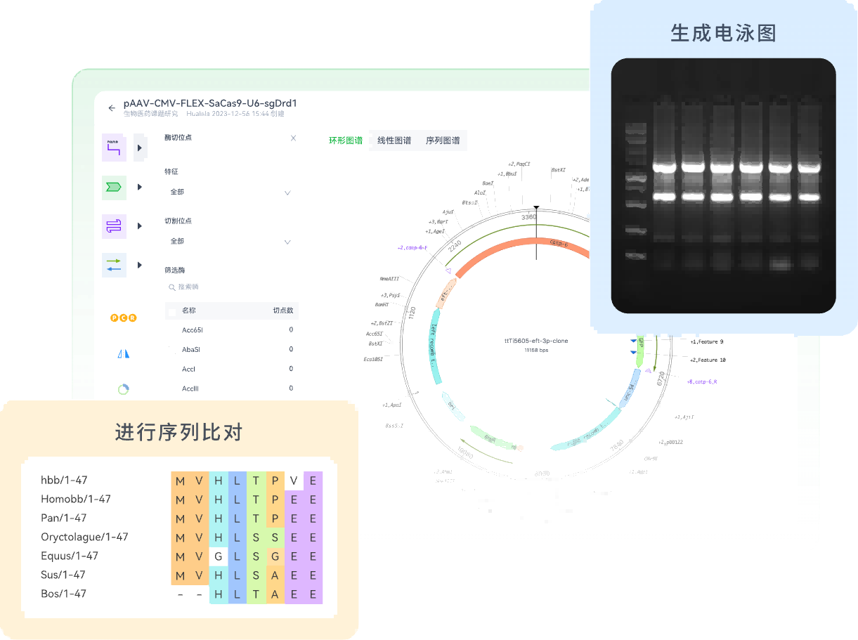
Simply performing a basic text comparison is inadequate for rigorous scientific quality control. Raw sequencing traces contain dye blob artifacts, baseline noise, and signal degradation at read termini that require chromatogram deconvolution. Establishing standardized pairwise and multiple sequence alignment workflows ensures that molecular biology teams catch cloning errors early and maintain complete sequence traceability.
Core Alignment Algorithms in Molecular Biology Quality Control
Different alignment algorithms serve distinct analytical roles in recombinant DNA verification:
1. Global Alignment (Needleman-Wunsch Algorithm): Global alignment forces an end-to-end comparison spanning the entire length of both sequences, maximizing matching scores across all positions. It is ideal for comparing two fully defined synthetic genes or homologous construct versions of similar length.
2. Local Alignment (Smith-Waterman / BLAST Algorithm): Local alignment identifies regions of high local sequence similarity, ignoring divergent or unmatched flanking regions. It is the gold standard for aligning individual Sanger sequencing reads (600–900 bp) against a large 5–12 kb plasmid backbone, identifying where the sequencing primer bound and checking insert junctions.
3. Multiple Sequence Alignment (MSA - Clustal Omega / MUSCLE): MSA aligns three or more sequences simultaneously. It is essential when aligning overlapping forward and reverse Sanger sequencing reads across an entire open reading frame to generate a high-confidence consensus contig.
4. Chromatogram Trace Deconvolution: Aligning raw .ab1 or .scf capillary trace files requires inspecting underlying fluorescent peak shapes. Double peaks at a single nucleotide coordinate indicate heterozygous mutations or mixed bacterial colony contamination.
Comparison of Sequence Alignment Methodologies
The table below summarizes common sequence alignment workflows used in molecular biology laboratories:
| Alignment Workflow Model | Algorithm & Scope | Chromatogram Inspection | Team Collaboration & Traceability | Best Fit QC Task |
|---|---|---|---|---|
| Free Online Text BLAST | Local heuristic alignment; compares raw FASTA text strings | None; cannot inspect underlying fluorescent chromatogram peaks | Zero record linking; results exist only in temporary browser sessions | Quick sequence identity spot-checks and species verification |
| Desktop Sequence Software (e.g., SnapGene, Geneious) | Pairwise and local alignment with manual chromatogram trimming | High; interactive electropherogram trace viewer with confidence bars | Isolated; files stored locally on individual user workstations | Single-user desktop clone screening and manual trace inspection |
| Connected Cloud Molecular Platform (e.g., Zettalab ZettaGene) | Automated multi-read alignment against vector reference with consensus contig generation | Full interactive trace viewer, automated low-quality end trimming, and variant flagging | Native entity linkage to master plasmid registries and ELN experiment records | Biotech teams, core facilities, and collaborative construct pipelines |
Step-by-Step Reference Alignment Protocol
To establish a reproducible sequence verification SOP, laboratories should execute a four-stage alignment workflow:
Step 1: Ingest Reference Map and Raw Trace Files: Import the verified in silico plasmid map containing full feature annotations. Upload incoming raw capillary sequencing files (.ab1/.scf) directly from the sequencing core or commercial vendor.
Step 2: Automated Quality Trimming: Apply automated quality clipping to remove low-confidence primer-binding regions (first 20–40 bp) and noisy, degraded 3' termini where signal-to-noise ratios drop below acceptable thresholds (Phred score < Q20).
Step 3: Align and Assemble Multi-Read Contigs: Align forward and reverse reads against the reference sequence. The software builds a merged consensus contig, highlighting any single nucleotide polymorphisms (SNPs), insertions, or deletions against the reference.
Step 4: Inspect Discrepancies and Document QC Sign-Off: Manually inspect any flagged mismatch against the original chromatogram trace. If the peak is clean and sharp in both directions, confirm the mutation; if the trace shows baseline noise or ambiguous overlapping peaks, order a re-sequencing run. Commit the validated alignment report to the electronic lab notebook.
Connecting Sequence Alignment to Laboratory Notebooks
When sequence alignment results remain trapped in isolated desktop files, bench scientists frequently rely on verbal confirmations, leaving experiment records without empirical trace evidence.
Within Zettalab, ZettaGene provides an integrated cloud sequence alignment environment. Molecular biologists can align raw ABI trace files or NGS consensus assemblies directly against reference plasmid maps, inspect interactive chromatograms, and embed visual alignment summaries directly into ZettaNote experiment records. This unified architecture ensures that construct design, sequencing QC data, and downstream functional assay results remain permanently synchronized.
FAQ
What is a Phred quality score in DNA sequencing alignment?
A Phred quality score (Q-score) is a logarithmic measure of base-calling error probability. A Q20 score corresponds to a 1 in 100 error rate (99% accuracy), while a Q30 score indicates a 1 in 1,000 error rate (99.9% accuracy). Bases with Q-scores below 20 should be trimmed before sequence alignment.
Why do the first 20 to 40 bases of a Sanger sequencing trace often show errors?
At the start of capillary electrophoresis, short primer-dimer fragments and unincorporated fluorescent dye terminators ("dye blobs") migrate rapidly through the capillary, generating broad fluorescent spikes that obscure true base calls. Automated quality trimming removes this initial noisy window.
How can researchers differentiate a true point mutation from a sequencing artifact?
A true point mutation displays a clean, symmetrical, single-color fluorescent peak that aligns with surrounding peak spacing and is confirmed in both forward and reverse sequencing reads. A sequencing artifact typically shows broad, asymmetrical, overlapping peaks with high baseline noise in only one read direction.
Can cloud-based sequence alignment tools handle whole-plasmid NGS consensus data?
Yes. Modern cloud molecular biology platforms import multi-kilobase consensus FASTA files from Nanopore or PacBio whole-plasmid sequencing runs, aligning the full circular sequence against the reference vector map to verify backbone and insert integrity in a single view.
Conclusion
Aligning DNA sequences against an annotated reference plasmid is a critical quality control milestone in molecular biology, ensuring that recombinant constructs match their intended design with single-base precision. By pairing automated quality trimming with interactive chromatogram inspection and integrated notebook records, research teams maintain high experimental standards. Explore Zettalab to align, analyze, and document your DNA sequencing datasets in a collaborative cloud workspace.
Related Articles